Electron Microscopy Service
The University of Navarra offers this service to those companies, universities and centers of research that request it.
AN ADVANCED ELECTRON MICROSCOPE THAT ALLOWS FOR ULTRASTRUCTURAL ULTRASTRUCTURAL ANALYSIS AT HIGH RESOLUTION
The electron microscope, which combines the main functions of a scanning electron microscope (SEM or Scanning Electron Microscope) and additionally has a transmitted electron detector (STEM or Scanning Transmission Electron Microscope), makes it possible to construct a three-dimensional image through the radiation emitted by a surface after being bombarded with electrons, as well as to determine the composition of any subject from sample. One of the main differences between this equipment and other more conventional ones is the magnification scale and the different types of compositional analysis possible.
Chemistry
+
In the chemical field, the different Materials (ceramics, metals, polymers, etc.) are studied and in the pharmaceutical industry it is possible to study nanoparticles, etc.


Biology
+
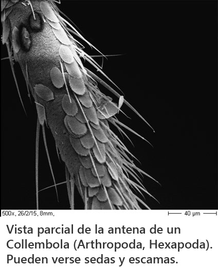
On the area of Biology it can be used to see small organisms, and also their parts, even cells or the surface of tissues.

Biomedicine
+
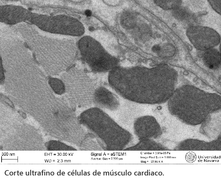
In Biomedicine, it enables the ultrastructural analysis of normal and pathological cells and tissues. In some diseases it allows the detailed study of aspects such as mitochondrial damage, autophagy processes, presence of protein aggregates, as well as the characterization of extracellular vesicles and viruses.

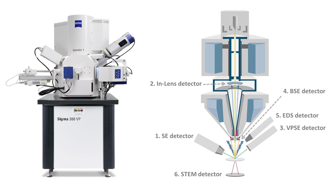
The FE-SEM, using two different detectors (SE2, inLens), offers the possibility to observe samples at a magnification of up to 300,000x. High resolution images can be achieved, with a limit of less than 1 nm (nm = nanometer; 1 nm = 0.000001 mm). The equipment accepts samples up to 1 cm in diameter, and ideally they should not be very tall (up to 5 mm). Flat sample holders (stub) of 12.7 mm diameter are usually used. They must be completely dehydrated when introduced into the vacuum chamber, but there are no limitations as regards their composition: cells, tissues, small organs, small animals, pieces of plants, and of course any object of a plastic or metallic nature.
The service is complemented with a "critical point" drying equipment, which allows dehydration of any object through an intermediate alcohol-CO2 step using a pressure chamber; afterwards it is convenient to deposit a thin layer of gold (16 nm) using a shader (we have a Sputter-Coater brand Emitech Ltd., model K550, Strovolos, Cyprus).
data technicians
→ Schottky thermal field emitter.
→ A 30 kV (STEM): 1.0 nm
→ A 30 kV (VP mode): 2.0 nm
→ A 30 kV (VP mode): 2.0 nm
→ At 1 kV: 1.6 nm (2.2 nm Emitter Thermal field emission gun (FEG), stability > 0.2 %/h)
→ 50 ns/pixel
→ 0.02 - 30 kV
→ 10× - 1,000,000×
→ 8.5 mm cut and a take-off angle of 35°.
→ 3 pA - 20 nA 100 nA optional (4 pA - 20 nA; 40 nA - 100 nA at high current)
RATE OF THE TECHNIQUE |
||
| Gold coating | 36,00 € | Up to 6 samples |
| Negative contrast - no osmium | 50,00 € | Up to 4 samples |
| Negative contrast - with osmium | 70,00 € | Up to 4 samples |
| Fixing and embedding in resin | 80,00 € | Up to 4 fragments of 1 sample |
| Fixation, agar pre-embedding and resin embedding | 100,00 € | Up to 4 fragments of 1 sample |
| Semifine slices stained with toluidine blue | 40,00 € | Up to 2 fragments of 1 sample |
| Cutting and contrasting grids | 48,00 € | 2 grids per fragment |
| Additional grids of the same fragment | 20,00 € | 1 grid |
| Immunohistochemistry (without primary antibody) on semi-thin slices | 15,00 € | Per slide |
| Immunohistochemistry (without primary antibody) on grids | 25,00 € | Per grid |
| Immunohistochemistry (without primary antibody) and negative contrast - without osmium | 150,00 € | Up to 4 samples |
MICROSCOPE USE RATE PER HOUR * |
||
| Users University of Navarra and CIMA | 60,00€ | |
| Agents of SINAI (Navarre System of research and development+i) | 87,00 € | |
| External users | 153,00 € | |
* VAT not included
application of the service
In this document reflects important information about the processing of the samples prior to their observation. Before filling out the form it is essential to read this information. The requests will be sent to the persons in charge of each area and they will contact contact to confirm the budget and to specify a quotation to carry out the essay.
Chemistry
Adrian Duran
948 425 600 (Ext. 806647)
adrianduran@unav.es
Environmental Biology
Enrique Baquero
948 425 600 (Ext. 806495)
ebaquero@unav.es
Biomedicine
Marián Burrell
948 425 600 (Ext. 806247)
mburrell@unav.es


![]()


![]()
