The University acquires an electron microscope for the area of Sciences.
The new equipment will be used for the teaching, to improve the research, and to provide services to companies.
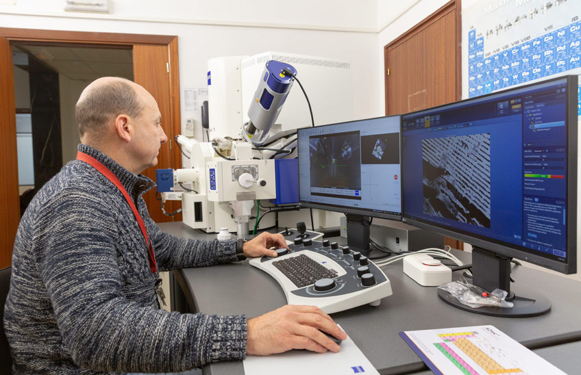
FotoManuel Castells<br>/El profesor Enrique Baquero, trabajando con el nuevo microscopio electrónico.
25 | 01 | 2023
The area of Sciences of the University of Navarra has a new Scanning Electron Microscope (FESEM) since last November. This equipment will be used to carry out research in the Schools of the area of Sciences, will be used for the teaching, and will also provide service to companies and other universities and centers of research that request it.
The electron microscope, which combines the main functions of a scanningelectron microscope (SEM or Scanning Electron Microscope) and additionally has a detector of transmitted electrons(STEM or Scanning Transmission Electron Microscope), allows the construction of a three-dimensional image through the radiation emitted by a surface after being bombarded with electrons, as well as allowing to know the composition of any subject of sample.
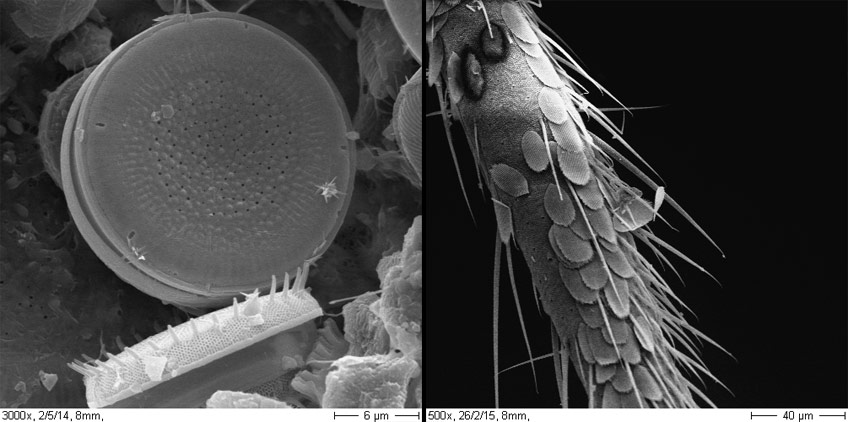
Microscope samples of a diatom algae and part of an insect antenna.
"In the field of research this microscope allows us to illustrate our scientific publications with high magnification and high quality photos of living beings, objects and materials" says Enrique Baquero, director of department of Environmental Biology and one of the researchers responsible for the new microscope.
In the field of Chemistry and materials science, the study of polymers, nanoparticles, ceramics, etc. is possible. In the biologicalarea it is possible to see life forms (e.g. very small animals) or their parts - even cells or tissues - and in medicine it will allow to answer some questions, for example, in detailed programs of study of mitochondrial damage, of the autophagy process, the characterization of extracellular vesicles and of viruses, etc.
One of the main differences of this equipment with respect to other more conventional ones is the magnification scale and the different types of compositional analysis possible. "The equipment can perform quantitative elemental chemical analysis at one point of the sample, in addition to giving stratigraphic composition profiles and compositional mapping of different zones of the materials analyzed" points out Adrián Durán, professor at department of Chemistry and one of the professors responsible for this microscope.
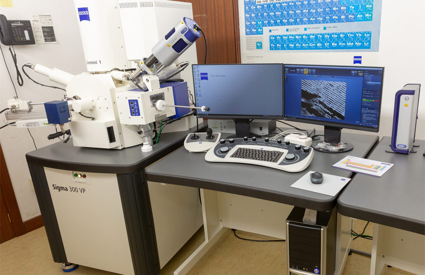
The scanning electron microscope (FESEM).
The microscope is located on the third floor of Research Building.
This microscope, which cost around half a million euros, is 50% subsidized by the Government of Navarra's call for infrastructures.