Automatic cell tracking to identify alterations involved in cancer
Dr. Carlos Ortiz de Solórzano, director of the Imaging Platform at CIMA, coordinates an international competition that analyzes 52 microscopy videos.
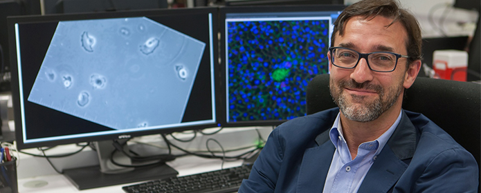
"By studying the movement and proliferation of cells, we can learn how living organisms function, both in normal processes and in alterations involved in diseases such as cancer," explains Dr. Carlos Ortiz de Solórzano, director of the Imaging Platform and laboratory of Preclinical Models and Analysis Tools at research center Applied Medicine (CIMA) of the University of Navarra. The researcher coordinates an international challenge or competition that analyzes 52 microscopy videos. The committee organizer of the challenge involves the researcher Arrate Muñoz Barrutia, from the Carlos III University of Madrid, together with scientists from the Czech Republic, the Netherlands and Germany. The results have been published in the latest issue of Nature Methods, a specialized journal of high scientific impact.
Many cells of living beings must migrate to fulfill their functions, both during the embryonic development as well as in the adult individual. It occurs in normal processes and in abnormal processes such as cancer, when they leave the primary tumor to generate metastasis and colonize other tissues. "Studying how cells move financial aid to learn about these normal and pathological processes. tool But cell tracking is also a very useful tool to know their genealogy, that is, where the cells of an organ come from, so that we can study the initial processes of a disease".
92 GB from data characterized and annotatedThe competition presented in Nature Methods compiles the results of the three editions of the challenge, in which 21 groups from 18 countries participated, on cell detection and tracking in two- and three-dimensional microscopy videos. In total, 52 videos were analyzed, occupying 92 GB. Some are synthetic videos, created by a cell simulator, and the rest are real, from brightfield microscopy, fluorescence, bidirectional, three-dimensional...
According to researcher of CIMA, who is also director of Master's Degree in Biomedical Engineering at Tecnun and member of research center at network in Oncology (CIBERONC), "this work performs an analysis of the algorithms used for tracking the videos included in the competition. The conclusions of this analysis provide very interesting information for software developers, since we have a very varied, characterized and annotated data . In addition, it provides information of interest also for potential users of these programs. In particular, we have found that algorithms that use learning techniques and those that perform tracking as a whole, considering the entire life of the cells, rather than performing temporal associations close in time, work better".
Dr. Ortiz de Solórzano recognizes that "there is still a lot to do. The challenge is still open online and the next step is to add new data, especially those with a more complex analysis due to their Issue, such as the videos that capture the embryonic development of living beings".
reference letter bibliographic
V. Ulman, M. Maska, several authors et. C. Ortiz-de-Solorzano. An objective comparison of cell-tracking algorithms. Nature Methods (2017). Advance online Publication doi:10.1038/nmeth.4473.